Yuewen Peng1,
Bin Yu1 ![]() ,
Fang Liu2
,
Fang Liu2
For correspondence:- Bin Yu Email: yu_bin2000@sina.com Tel:+862061641741
Received: 19 May 2015 Accepted: 28 December 2015 Published: 28 February 2016
Citation: Peng Y, Yu B, Liu F. Epigallocatechin-3-gallate promotes osteoblastic activity in human osteoblast-like cells. Trop J Pharm Res 2016; 15(2):313-317 doi: 10.4314/tjpr.v15i2.13
© 2016 The authors.
This is an Open Access article that uses a funding model which does not charge readers or their institutions for access and distributed under the terms of the Creative Commons Attribution License (http://creativecommons.org/licenses/by/4.0) and the Budapest Open Access Initiative (http://www.budapestopenaccessinitiative.org/read), which permit unrestricted use, distribution, and reproduction in any medium, provided the original work is properly credited..
Purpose: To investigate the effect of epigallocatechin-3-gallate (EGCG) on bone metabolism and osteoblastic activity.
Methods: MG-63 human osteoblast-like cells were treated with varied concentrations of EGCG. Alkaline phosphatase (ALP) activity and matrix mineralization assays were carried out on the treated and untreated MG-63 human osteoblast-like cells. Beta-catenin mRNA level was determined by quantitative real-time polymerase chain reaction (q-PCR).
Results: The results showed that EGCG treatment significantly increased ALP and mineralization activities at concentrations of 15 and 30 μM, in a dose-dependent manner. Furthermore, EGCG treatment significantly increased beta-catenin mRNA ex
Conclusion: EGCG promotes osteoblastic activity in human osteoblast-like cells, by Wnt signaling through estrogen receptor (ER) pathway.
Introduction
Osteoporosis is a musculoskeletal disease characterized by low bone mineral density (BMD) and high risk of fragility fractures. Osteoporosis has drawn more and more attention from medical researchers because it is becoming an increasing risk for human health and quality of life [1]. Osteoporosis is caused by imbalance between bone formation and bone resorption, which relies on the interactions between osteoblasts and osteoclasts [2,3]. Osteoblasts, known as bone-forming cells, are the major osteoprogenitor cells, whose proliferation and differentiation will eventually result in the formation of the mineralized extracellular matrix [4].
To date, one effective solution to treat osteoporosis is targeted on osteoblasts by increasing the proliferation of the osteoblastic lineage and promoting the differentiation of osteoblasts, and eventually to induce new bone formation [5]. The functions of osteoblasts can be regulated by various local and hormonal factors such as Wnt glycoproteins. Canonical Wnt/beta-catenin signaling pathway plays an important role in the maintenance of bone mass and bone formation [6]. Wnt ligands binding to the frizzled receptor will lead to accumulation and stabilization of beta-catenin in the cytoplasm [7]. Recent studies demonstrate that beta-catenin through Wnt signaling is essential in skeletal development through the regulation of osteoblastogenesis [7,8].
Epigallocatechin-3-gallate (EGCG), a component of green tea, has been demonstrated to possess various biological activities, including anti-cancer, anti-inflammation, anti-bacteria and so on [9-11]. In the present study, we studied the effects of EGCG on bone metabolism regarding osteoblastic activity in MG-63 human osteoblast-like cells, and evaluated the possible underlying mechanisms.
Methods
Cell culture and cell viability assay
Human osteoblast-like MG-63 cells were obtained from the Institute of Biochemistry and Cell Biology, the Chinese Academy of Science, Shanghai, China. The cells were cultured in Dulbecco’s modified Eagle’s medium (DMEM; Life Technologies, CA, USA), containing 10 % fetal bovine serum (FBS; Life Technologies, CA, USA) and 1 % penicillin and streptomycin (PS; Life Technologies, CA, USA) in a humidified incubator at 37 °C with 5 % CO2. Cell viability was determined by the 3-[4, 5-dimethylthiazol-2-yl]-2, 5-diphenyltetrazolium bromide (MTT; Sigma, St. Louis, MO). MG-63 cells were plated in 96-well plates (5 × 10³ cells/well) and treated with various concentrations of EGCG (7.5 – 30 μM) for three days. MTT solution at a concentration of 5 mg/mL was added into each well and incubated for another 2 h. The optical density [12] at 540 nm was measured and expressed as the percentage of vehicle control.
Alkaline phosphatase (ALP) activity assay
ALP activity was determined using a commercial Alkaline Phosphatase Microwell Substrate System (Sigma, St. Louis, MO) following the manufacturer’s instruction. After treatment with various concentrations of EGCG (7.5 – 30 μM) for 4 days, the cell monolayer was gently rinsed twice with ice cold PBS and lysed with lysis buffer containing 0.1 % Triton X-100, 50mM Tris-HCl (pH 7.2), and 2 mM MgCl2. The supernatant was collected after the lysate was centrifuged at 14,000 xg for 10 min at 4 °C. ALP activity in the clear supernatant was measured with ALP activity assay kit and the total protein concentration was determined with a BCA-protein assay kit.
Matrix mineralization assay
The degree of matrix mineralization of MG-63 cells was determined by Alizarin Red S staining method after EGCG treatment for 6 days [13]. Cells were washed twice with cold PBS and fixed with cold 70 % ethanol (v/v) for 30 min. Next, the cells were rinsed with distilled water and exposed to 40 mM Alizarin Red S solution (pH 4.2) for 10 min. After removing the unbonded stain by rinsing three times with distilled water, the amount of stain on the tested cells was extracted by shaking the cells with 10 mM sodium phosphate containing 10 % cetylpyridinium chloride (Sigma, USA) for 15 min. The values were then determined at an optical density of 562 nm. The data were expressed as percentage relative to the vehicle control group, after the relative matrix mineralization values were normalized by the relative cell viability.
NA extraction and quantitative real-time polymerase chain reaction (q-PCR)
The expression of beta-catenin mRNA was evaluated by q-PCR. The total RNA was extracted with TRIzol® reagent (Life Technologies, NY, USA), according to the manufacturer’s instructions. In the experiment, 1 μg RNA was used for the reverse transcription reaction using Oligo dT (18T) (Omega, NY, USA). Nucleotide sequences of primer for beta-catenin were: forward, 5’-gccacaggattacaagaagc-3’; reverse, 5’-ccaccagagtgaaaagaacg-3’.
In our experiment, PCR was carried out in duplicate with SYBR Green PCR Master Mix using a 7900HT qPCR system thermal cycler (Applied Biosystems, CA). Beta-actin mRNA was used as internal control for each sample. The Ct values for each sample were normalized to beta-actin mRNA. Nucleotide sequences of primers for beta-actin were: forward, 5’-ttctacaatga gctgcgtgtg-3’, reverse, 5’-ggggtgttgaaggtctcaaa-3’. Results were shown for three independent experiments.
Statistical analysis
The data were shown as the mean ± standard deviation from three independent tests. One-way analysis of variance (ANOVA) followed by Dunnett test was performed using Graph Pad Prism software version 5.0 for statistical analysis. In all experiments, p < 0.05 was considered statistically significant.
Results
Effects of EGCG on cell viability of MG-63 human osteoblast-like cells
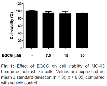
After treatment with various concentrations (7.5 – 30 μM) of EGCG for 3 days, cell viability was determined by the MTT assay. The data showed that EGCG did not affect the cell viability of MG-63 cells at the tested concentrations (), indicating that EGCG was not cytotoxic to MG-63 human osteoblast-like cells.
Effects of EGCG on ALP activity of MG-63 human osteoblast-like cells
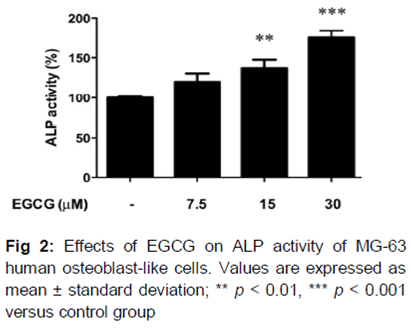
In order to determine whether EGCG could increase osteoblastic cell differentiation, its effect on ALP activity as investigated. Our results showed that treatment of MG-63 cells with EGCG for 4 days dose-dependently stimulated ALP activity in the cells. ALP activity was remarkably increased by 37.1 ± 10.6 and 75.6 ± 9.5 % with EGCG treatment at 15 and 30 μM, respectively, when compared with the vehicle control group ().
Effect of EGCG on mineralization activity of MG-63 human osteoblast-like cells
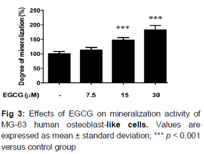
The matrix mineralization assay was also evaluated for further studying the effects of EGCG on osteoblastic activity. The data demonstrated that EGCG treatment also significantly promoted the matrix mineralization at 15 and 30 μM, by 47.3 ± 9.1 and 82.7 ± 15.2 %, respectively ().
Effect of EGCG on beta-catenin mRNA expression in MG-63 human osteoblast-like cells
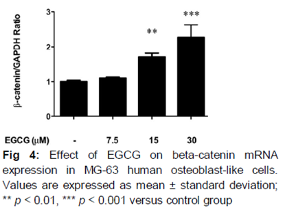
Beta-catenin is an important molecule for Wnt signaling pathway, which plays a crucial role in bone formation. Therefore, to determine whether Wnt signaling pathway was involved in the effects of EGCG on osteoblastic activity, beta-catenin mRNA expression levels in MG-63 cells were evaluated after EGCG treatment. As shown in , EGCG treatment significantly increased the beta-catenin mRNA expression. The beta-catenin mRNA levels were increased by 70.7 ± 11.0 and 126.7 ± 35.1 %, respectively by EGCG treatment at 15 and 30 μM.
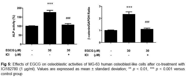
Effects of EGCG on osteoblastic activities in MG-63 human osteoblast-like cells after co-treatment with ICI182780
To determine whether estrogen receptor (ER) was involved in EGCG activated osteoblastic activities, ICI 182,780, an antagonist of ER was co-treated with EGCG on MG-63 cells. Our data showed that the stimulative effects of EGCG treatment on ALP activity were abolished by ICI 182,780 (1 μg/ml) co-treatment. In addition, the increasing mRNA level of beta-catenin by EGCG treatment was also inhibited by ICI 182,780 co-treatment ().
Discussion
In the present study, EGCG was found to be capable of increasing ALP activity and mineralization activity, in a dose-dependent manner. Besides, EGCG treatment significantly increased after beta-catenin mRNA expression by 70.7 ± 11.0 and 126.7 ± 35.1 %, respectively by EGCG treatment at 15 and 30 μM. Furthermore, the stimulating effects of EGCG treatment on ALP activity were abolished by co-treatment with ICI 182,780, an antagonist of estrogen receptor (ER). In addition, the increasing mRNA level of beta-catenin by EGCG treatment was also inhibited by ICI 182,780 co-treatment.
Osteoblasts play an important role in the process of osteoporosis. Among various osteoblast differentiation markers, ALP is known as a representative marker for the early stage of differentiation, and is a critical indicator of the initiation of mineralization during bone formation [2]. In our results, EGCG could significantly increase ALP activity and also the mineralization activity, indicating that EGCG has great potential in promoting osteoblastic activities.
Wnt/beta-catenin pathway plays an essential role in bone mass accrual, regulation and maintenance [12]. Scientific evidence reveals that beta-catenin is an important mediator in the Wnt signaling pathway, which plays a crucial role in bone formation [14]. Researchers also find that deletion of the beta-catenin gene will inhibit the proliferation and differentiation of osteoblasts [15]. In the current study, EGCG significantly increased the beta-catenin mRNA in MG-63 cells, indicating that the Wnt/beta-catenin pathway is involved in the actions of EGCG in osteoblasts. ER also plays a significant role the proliferation and differentiation of osteoblasts, and subsequently bone remodeling and development of the skeleton [16].
Clinical data show that human ER genes are positively related with femoral fracture and bone metabolism [17]. In our study, blocking ER with the antagonist ICI 182,780 also inhibited the beneficial effects of EGCG in MG-63, suggesting ER pathway is also involved in EGCG-induced osteoblastic activities.
Conclusion
Taken together, the findings demonstrate that EGCG promotes osteoblastic activity in human osteoblast-like cells, which may involve Wnt signaling through ER pathway. All the findings strongly suggest that EGCG needs to be further studied with a view to developing an effective therapeutic agent for osteoporosis.
References
Archives


News Updates